Dental and Beyond 972-437-0200

  CBCT

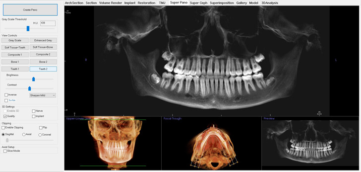
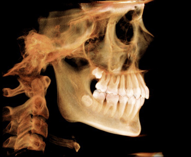
The in-office cone beam CT scanner is the state of the art from Imaging Sciences International. This brings the latest technology to dental diagnosis and treatment plans, yet the dose of radiation is extremely low.

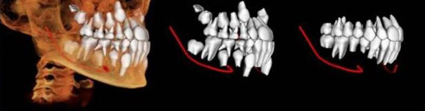
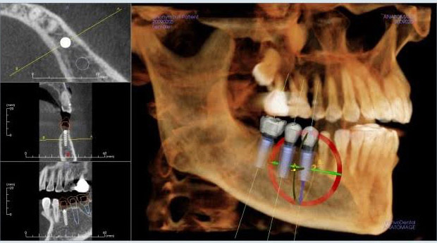
We can now measure exact bone width, thickness, height, and density for implant placement. Diagnosis of abnormalities in the bony structures is also possible to a degree not previously available. The relationship between the root of an impacted wisdom tooth or the exact location of any impacted tooth can now be determined instantly without guesswork.

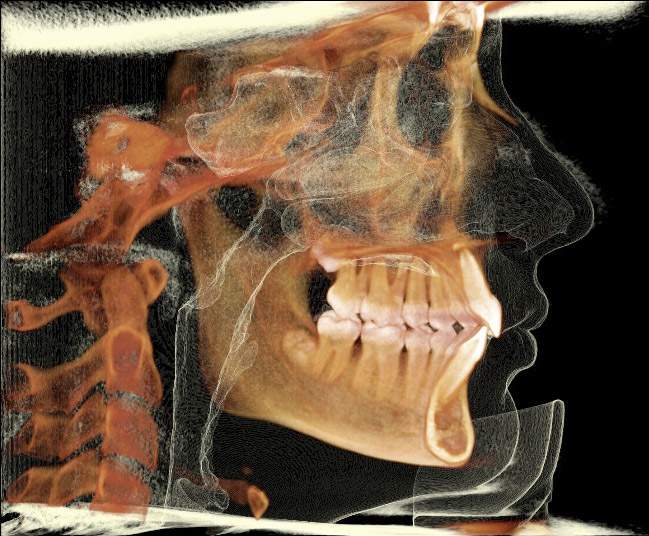
The following images show the amazing 3-D visuals now routinely possible in our office.

Cone Beam CT
Panoramic
CT Overview
3 views of teeth
CT No Overlay
Side view
CT Overlay
Soft tissue overlay
CT Overview
Detailed dimensions
CT Overview
After completion of 3 implants
3-D rotation around the back
3-D rotation in the front

  Implant

Case 1: This basic case restores a single missing tooth.

Before Implant
(1) Pre-treatment: dead tooth
Extraction
(2) Hopeless tooth extracted
Single Implant Placed
(3) Implant placed after healing
Abutment Inserted
(4) Abutment inserted into implant
Single Implant
(5) Crown made on top of abutment
Completed Implant
(6) Final appearance in mouth

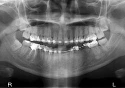
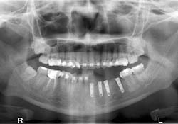
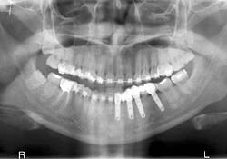
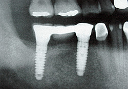
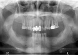
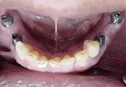
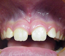
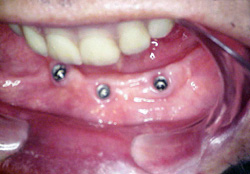
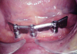
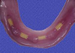
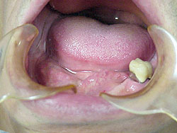
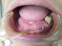
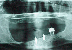
Case 2: Multiple missing teeth are restored by implants. Orthodontics are first used to align the teeth properly. Dentures over the implants are used to replace the missing teeth.

Braces Before Implants
Braces around missing teeth
Braces Implants Placed
Implants placed after orthodontics
Braces Implant After
Implant-supported fixed dentures

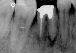
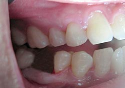
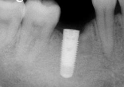
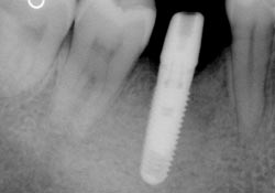
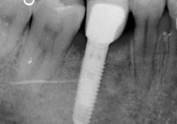
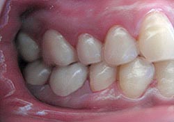
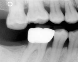
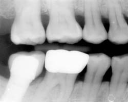
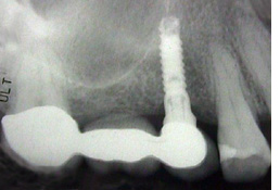
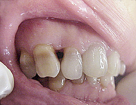
Case 3: An implant replaces a missing tooth and treats a super-erupted tooth.

Super-eruption Before
Molar (top) has super-erupted due to missing opposing tooth (bottom)
Super-eruption After
Super-erupted molar intruded after implant placed

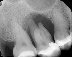
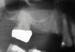
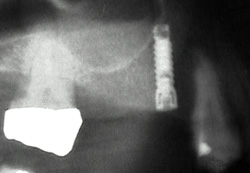
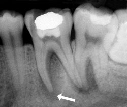
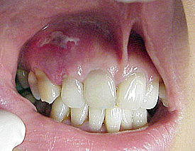
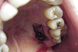
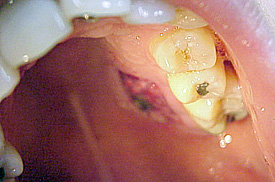
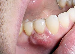
Case 4: A severely infected tooth with severe bone loss underwent bone graft and sinus lift to enable implant.

Bone Graft Before
Infected molar (middle) surrounded by pus and bone loss
Super-eruption After
Healthy bone restored for implant, after bone graft, sinus lift

  Orthodontics

Case 1: This rather complex case shows multiple defects to be corrected in this child.

Cast Before
Cast: severe overbite and crowding
Alignment Before
Alignment before intervention
Appliance in Place
Appliances in place for 1 month
Alignment After
Removal of appliances after 18 months

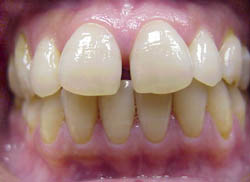
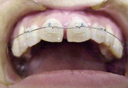
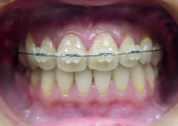
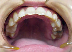
Case 2: Sectional orthodontics refers to a quick, smaller scale intervention, as shown in this adult case.

Gap Before
Prominent gap before intervention
4 Brackets
Placement of only 4 brackets
Gap After
Status of our patient after 1 month
Removal Gap
Removal of appliances after 3 months

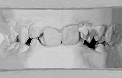
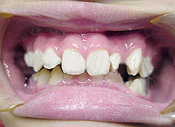
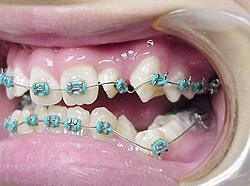
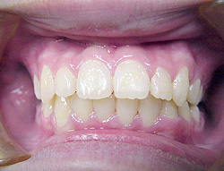
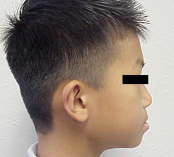
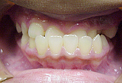
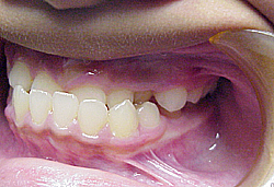
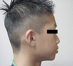
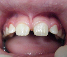
Case 3: Here is a case showing repair of a severe underbite in a child.

Underbite Before
(1) Profile showing protruding lower jaw
Underbite After
(4) Improved profile after 15 months
Anterior After
(5) Our orthodontic treatment result
Tilt After
(6) Correction of underbite without surgery

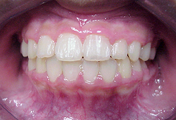
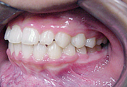
Case 4: Orthodontic treatment can improve narrow airway spaces, as shown in this teenager.
The patient played tennis but was limited by fatigue prior to orthodontic treatment.
After correction she was able to last longer in tennis and play her full game.

Airway Before
Severe overbite with narrow airway before orthodontics
Volume Before
Volume analysis confirms anatomical limitation
Airway After
Normal bite achieved in fewer than 2 years
Volume After
Blue/green confirm improved airway

  Implant Bridges

Case 1: Implant bridge is placed for 2 or more missing teeth.

Before Implant
Before implant: 2 missing teeth
Single Implant
Single implant placed
Tooth Implant Bridge
Bridge supported by natural tooth and implant
Total Implant Bridge
Total implant-supported bridge

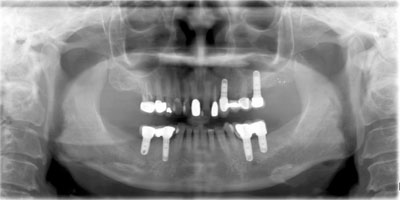
Case 2: A pair of implant bridges replace 6 missing teeth.

Implant Bridge Before
Missing teeth (bottom jaw)
Implants Placed
Implants placed (bottom jaw)
Implant Bridge After
Implant bridge in place
Implant Bridge Placed
Restored appearance and function

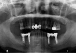
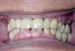
Case 3: This complex case involves a single tooth implant replacement, implant + natural tooth supported bridge, and left + right implant + implant supported bridges.

The Works
Multiple approaches for 1 patient

  Root Canal

Case 1: This shows a typical root canal treatment.

Before Root Canal
Dark shadow is infection in the bone around the 2 roots
After Root Canal
After root canal, the surrounding bone has healed

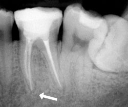
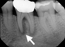
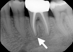
Case 2: This is another case showing the healing of the surrounding bone 1 year later.

Before Root Canal
Disease around the roots shows as a dark shadow in the bone
After Root Canal
The surrounding bone has healed 1 year later

  Oral Surgery

Frenectomy (removal of excess tissue that can pull on the gums, lips, or tongue) is an excellent example of a minor surgical procedure that can be done in our office. This case shows a child’s teeth.

Frenum Before
Tight frenum pulling on lips and teeth
Frenum Removed
Frenectomy to remove excess tissue
Frenum Final
Corrected appearance and function

  Implant Dentures

Case 1: This shows how all the components of an implant denture work together.

Implants Placed
Implants placed in toothless lower jaw
Denture Attachment
Completed attachment ready for denture
Denture Prosthesis
Inside of denture prosthesis with cushions
Implant Dentures After
Implant dentures in place (lower jaw)

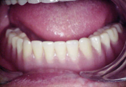
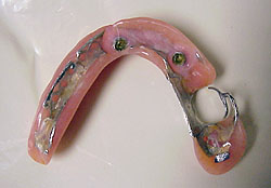
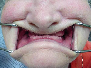
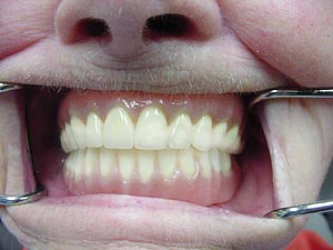
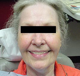
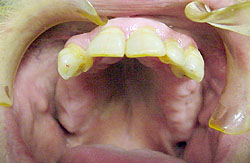
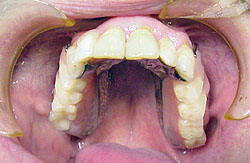
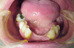
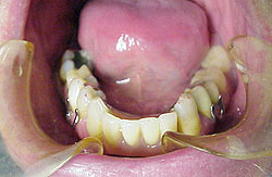
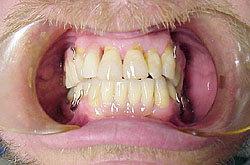
Case 2: Even a single tooth can be valuable as part of an implant denture solution.

Dentures Before
Edentulous except for 1 tooth
Attachment for Dentures
Implants with locking ball attachments
X-Ray
Implants in areas of severe bone loss
Denture Appliance
Appliance locks securely with 2 rings

  Partial Dentures

Case 1: Simple but important set of full dentures improves function but also appearance.

Without Dentures
(1) Edentulous mouth
Full Dentures
(4) Full dentures in place
Dentures After Face
(5) Restored facial appearance
Dentures After Profile
(6) Normal profile with full dentures

Case 2: Partial dentures are another basic but important tool.

Upper Teeth Before
Upper teeth before fitting
Upper Partial Dentures
Upper removable partial denture
Lower Teeth Before
Lower teeth before fitting
Lower Partial Dentures
Lower removable partial denture
Partial Dentures After
Total appearance with upper and lower partials in place

  Gum Graft

Case 1: This shows a soft tissue graft to correct a gum defect.

Gum Defect Before
Initial distorted gums with tissue defect
Gum Defect After
Corrected appearance 5 days after graft
Gum Harvest
Graft harvest site (roof of mouth)
Gum Healing
Site will continue to heal fully

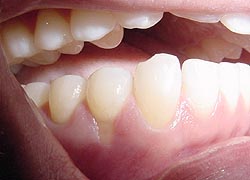
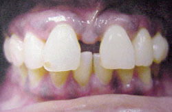
Case 2: Gum grafts are also important tools to cover exposed tooth roots.

Exposed Root
Receded gumline has left dangerously exposed tooth root
Graft Donor
Graft donor site after 1 week
Graft Take
Graft takes at 1 week
Donor Site Healed
Donor site healed at 2 weeks
Graft After
Final appearance after 2 weeks

Case 3: Here is a case showing repairing one (on the left) out of multiple exposed roots. The other root (the one on the right) was repaired later.

Exposed Roots Before
2 severely exposed tooth roots
Exposed Roots After
Exposure repaired by gum graft

  Cosmetics

Case 1: Cosmetics can rely on combinations of crowns, cleaning, bleaching, and of course our dental spa to achieve overall results.

Cosmetics Before
Pre-treatment: hopeless case?
Cosmetics After
Completion of the front teeth: not so hopeless!

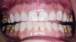
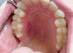
Case 2: Basic teeth cleaning again makes a dramatic improvement.

Cleaning Before
Bottom teeth prior to deep cleaning
Cleaning After
After much-needed cleaning... next, how about some bleaching?

  Bonding

Bonding is a quick solution to problems such as gapped teeth. This case shows a darkly pigmented individual.

Bonding Before
Significant gap between front teeth
Bonding After
Direct bonding completed in same office visit

  Crowns

Conventional crowns are one of the most basic procedures. (Now this patient just needs some alignment for the bottom teeth.)

Crown Before
Teeth prior to crown placement
Crown After
After crowns, as well as basic cleaning

  Bleaching

This frequently requested procedure is simple and effective. Note that temporarily even the surrounding gums can harmlessly take some bleaching effect. This case shows a darkly pigmented individual.

Bleaching Before
Unsightly stains prior to bleaching
Bleaching After
Same teeth after short period of bleaching

  Tooth Filling

Tooth-colored fillings are routine but can also be used to replace older fillings.

Filling Before
Unsightly old filling before replacing
Filling After
Same tooth with tooth-colored filling

  Oral Cancer

Sometimes suspicious lesions are discovered on dental exams. This is another important reason to have routine exams done.

Oral Lesion
Lesion we discovered on a routine check-up
Squamous Carcinoma
Diagnosis: squamous carcinoma (oral cancer)

  Dental Sleep Medicine

This is a normal patient who does not suffer from snoring or sleep apnea. The 3-D volume calculation shows normal volumes of the upper airway (white, at the far right of the color scale).

Normal View
Image slice showing mouth and airway
Normal 3D
White 3-D volume is normal

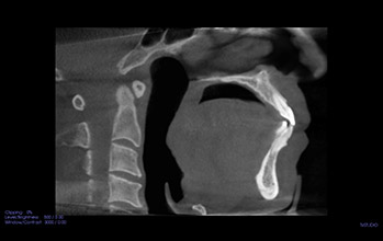
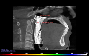
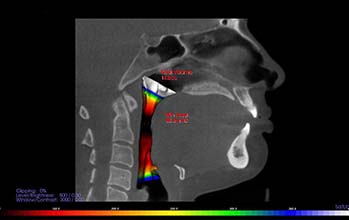
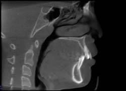
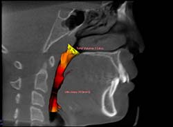
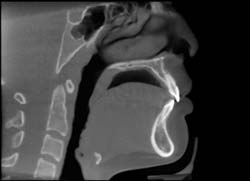
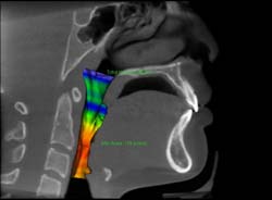
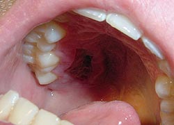
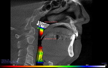
This patient suffers from loud snoring and has been diagnosed with obstructive sleep apnea. The 3-D volume calculation shows severe narrowing of the upper airway, which is causing these problems.

The middle of the calculated volume (an extremely important area!) shows the color gradient going all the way to black. Innovative oral devices can then be applied to solve the airway problem, including the snoring and drop in blood oxygen.

Constriction View
Image shows apparent narrowing
Constriction 3D
Black volume confirms extreme narrowing

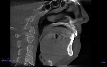
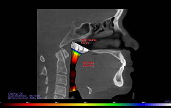
This patient came in complaining of poor sleep quality every night for many years. A night guard, a popular dental appliance, was suspected as a cause.

When wearing the night guard, the mid portion of the airway volume drops to hardly any (the color gradient blacking out). Without the night guard, the airway volume is still low but improves. The night guard was discontinued, resulting in improved sleep.

With Nightguard
Severe narrowing when wearing night guard
Without Nightguard
Reduced airway narrowing without night guard